Gastroscopy
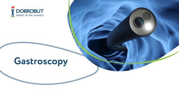
Gastroscopy or esophagogastroduodenoscopy is an examination of upper gastrointestinal (esophagus, stomach, duodenum) with the use of an endoscope. Gastroscopy allows to view gastrointestinal organs from inside, receive precise and full information and assess the condition of the mucous coat of upper gastrointestinal and also to trace dynamic changes after the surgery.
Light radiation in the special spectrum helps to determine possible changes on mucous membranes; additional staining of the mucous membrane also allows to diagnose organic changes, precancerous changes included.
Gastroenterologists have the possibility to develop an efficient treatment strategy which corresponds to international protocols relying on the results of lab tests data (if a check for Helicobacter pylori presence or a biopsy has been required).
Gastroscopy is prescribed under the following conditions:
- abdominal pains, nausea, vomiting, epigastric burning;
- food swallowing difficulty;
- complaints about postprandial fullness after eating the usual amount of food;
- complaints about pains in upper gastrointestinal with loss of appetite, abrupt body weight loss, anemia;
- the examination is recommended for the people over 45.
If your family members have a history of stomach cancer, you also have indications for such an examination.
Gastroscopy allows to reveal neoformations and determine their nature histologically. As a result of this examination, malignant gatrointestinal tumors are revealed at early stages; no other diagnostic technique gives such a possibility.
Besides, gastroscopy allows experienced specialists to reveal the diseases which may cause cancer (oncology).
The procedure has no absolute contraindications. However, it should be prescribed carefully for the patients with perforation of abdominal organs and decompensated respiratory and cardiovascular diseases.
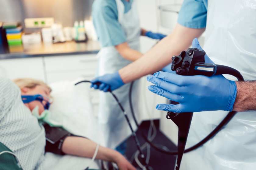
Description of gastroscopy
Preparation
Some preparatory measures should be taken for the scheduled gastroscopy.
- Exclude chocolate, nuts, seeds and spicy food from your menu, and you also must not drink alcohol.
- A light dinner is allowed in the evening before the procedure.
- The period of time between the last meal and the beginning of the procedure should be not less than 18 hours.
- You must not either eat nor drink 4–6 hours before the procedure and postpone medication taking.
A doctor applies anesthesia on the root of tongue and the pharynx with the spray just before the procedure. It allows to reduce a sense of discomfort during the procedure and also to reduce the presentation of the vomiting reflex. At Dobrobut clinic chain you can undergo the absolutely painless type of this diagnostic procedure using medication sleep.
The patient is located on a couch lying on the left side. A special device is inserted into the mouth that prevents the patient from occluding teeth during the procedure. The procedure itself includes the insertion of the endoscope, which is a thin tube with an optical device at the end, through mouth into esophagus, stomach, duodenum. During the checkup an endoscopist will gradually move the endoscope through the gastrointestinal tract with its parallel examination.
During the examination the specialist will be able to take a fragment of the mucous coat (to perform biopsy) to test it for the presence of Helicobacter pylori bacteria and to remove the polyps in the stomach or in the esophagus (if revealed) with further histopathological examination in a pathologic laboratory.
Our advantages
Cost of gastroscopy services
Choose the nearest clinic
ISO certificates
Accreditation certificates
Medical practice licenses




%402x.png)
%402x.png)
