MRI of the head
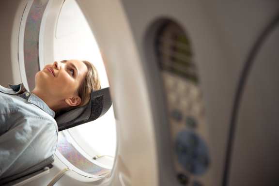
MRI of the brain today is one of the most effective and safe methods of neuroimaging, with which it is possible to assess the state of all structures of the brain (vessels, membranes, gray matter, as well as the bones of the skull), to determine the presence of neoplasms (tumors, cysts), to track the consequences of trauma, signs of inflammation or cerebrovascular accident. The examination is carried out using a tomograph - an apparatus that allows you to obtain a three-dimensional image of the areas under study. The resulting picture is shown on the screen and, if necessary, recorded on a CD. Within the framework of diagnostics, the body is not exposed to radiation exposure, which allows it to be carried out the required number of times without restrictions.
In certain cases, MRI with a contrast agent is used to view blood vessels and nerve endings. The study involves the intravenous administration of a drug based on gadolinium salts to the patient, which spreads through the vessels of the body and enhances signals from the tissues. The use of examination of the brain with contrast helps not only to clarify and detail the identified pathological changes but also in some cases to identify pathology that was not determined by a standard study.
Indications for MRI of the brain
- complaints of persistent headache;
- increased dizziness, fainting conditions;
- double vision;
- the presence of tinnitus;
- memory impairment decreased concentration;
- violations of coordination and orientation in space;
- numbness and weakness in the limbs;
- acute violation of cerebral circulation (stroke);
- tumor lesion of the brain and its membranes;
- diagnostics of demyelinating and degenerative diseases of the brain and assessment of the dynamics of changes during treatment;
- diagnostics of inflammatory diseases of the brain;
- pathologies of cerebral vessels (aneurysms, arteriovenous malformations, etc.);
- traumatic brain injury;
- as a check before and after surgery.
Despite the high safety of the procedure, it still has some contraindications.
Absolute contraindications include:
- the presence of intracranial ferromagnetic hemostatic clips of cerebral vessels;
- implanted electronic medical devices (implanted hearing aids, ferromagnetic and electronic middle ear implants, pacemakers, etc.);
- foreign metal objects in the soft tissues of the body (for example, shrapnel, bullets or fragments of bullets, metal shavings in the orbital area);
- heart valve prostheses;
- insulin pumps;
- weight exceeding the specifications of the tomograph;
- inability to stay still due to neurological or psychiatric illness.
Relative contraindications are:
- period of pregnancy;
- claustrophobia, which is accompanied by panic attacks;
- wearing non-removable braces for bite correction;
- the presence of tattoos on the body, with dyes on a metal basis (except for dyes based on titanium compounds).
- the presence of allergic reactions to the components that make up the contrast (iodine-containing drugs);
- renal failure (due to complicated excretion of contrast medium).
Preparing for the procedure
Conducting an MRI of the head does not require serious preparatory measures. If the patient has metal implants, pacemakers, artificial heart valves, any other foreign objects in his body, it is necessary to inform the doctor about this. Women should inform the specialist about the pregnancy. If the patient is shown to undergo an MRI of the brain with contrast, an allergic reaction to the contrast agent must be excluded in advance of the procedure by carrying out a tolerance test and blood tests for kidney tests.
Before the procedure, it is recommended to take off all metal and magnetic objects, including jewelry, hairpins, etc., as well as remove plastic cards, keys, telephones from your pockets. To minimize the risk of nausea and destabilization of blood pressure, it is recommended that the examination be performed on an empty stomach, within a few hours after the last meal. Smoking, coffee, and alcoholic beverages are not allowed before the procedure.
Procedure progress
The patient is placed on a special movable table, which slides into the tomograph and fixes the head. During the procedure, the patient must lie motionless. The clicks produced by the magnet help to understand that the shooting has begun. Communication between the patient and the doctor during the examination takes place using a speakerphone. On average, the diagnostic procedure takes about 30-40 minutes.
If the patient is shown to conduct an MRI of the brain with contrast, then the procedure is carried out in several stages:
- First, a traditional MRI procedure is performed without the use of contrast.
- Then the patient is injected into the vein with a contrast agent using a syringe system. Within 10-30 seconds, the drug spreads into all vessels and tissues and "highlights" the areas necessary for diagnosis.
- Then another MRI scan is performed.
- And finally, the preparation of the scan results is carried out, the study and description of the material by a radiologist.
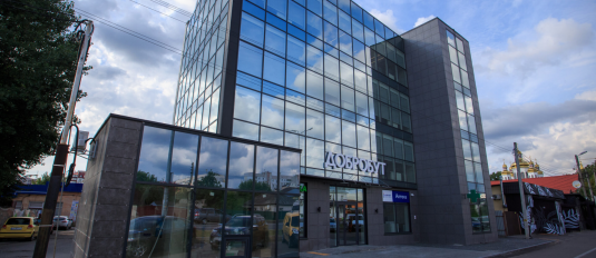
The Dobrobut network of medical clinics invites patients to conduct an MRI of the brain in Kyiv at affordable prices. Our center is equipped with the most advanced technologies in Ukraine, which helps to most accurately diagnose pathological changes. You can sign up for the procedure, as well as clarify its cost, by calling our call center.
Bibliography
- What is a head MRI? - Prattville Imaging Center Montgomery, AL- Medtech Imaging Center. Published online, 2020.
- MRI - Mayo Clinic. Published online, September 04, 2021.
- MRI scan. Published online, August 09, 2018.
- What to Expect in an MRI for the Head and the Brain | Envision Radiology. Published online, 2021.
- Magnetic Resonance Imaging (MRI) – Head. Last update February 08, 2021.
Our advantages
Our services
Choose the nearest clinic to you
ISO certificates
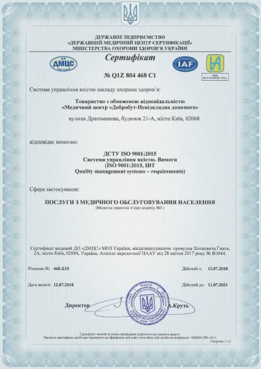
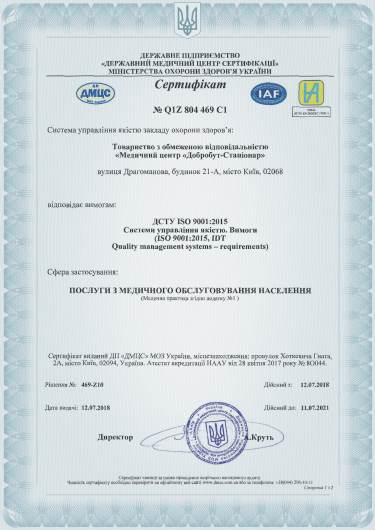
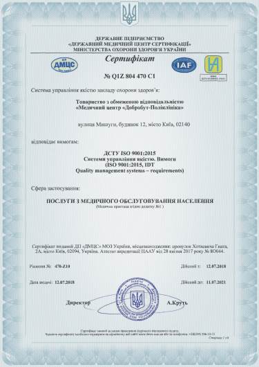
Accreditation certificates
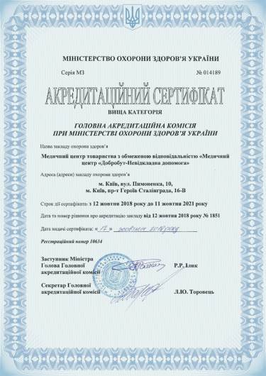
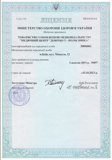
Medical practice licenses




@2x.png)
@2x.png)
%402x.png)
%402x.png)
