
Magnetic resonance imaging today is a relatively safe and high-precision diagnostic procedure that can determine the condition of soft tissues, abdominal organs, pelvis, joints, cartilage, intervertebral discs and brain. With the help of magnetic resonance imaging, namely radio frequency pulses and magnetic field, specialists obtain the clearest possible image of the patient's organs and tissues, which allows to detect even the initial changes of various nature in them.
Magnetic resonance imaging is performed in two ways:
- without contrast;
- with contrast .
Contrast-enhanced MRI is prescribed when it is necessary to obtain deeper characteristics of the process and is most often used in oncology to detect tumors in the early stages. And also to control the behavior of the tumor during treatment, as the method allows to detect the slightest changes.
The decision on how to perform MRI diagnosis (with or without contrast) is made by a radiologist based on the doctor's recommendations.


Equipment
The modern Toshiba Elan 1,5T MRI machine, which is used for diagnosis in the Dobrobut medical network, is designed for accurate diagnosis in the most comfortable conditions for the patient. Various programs built into this MRI machine increase the speed of the examination and facilitate diagnosis for medical staff.
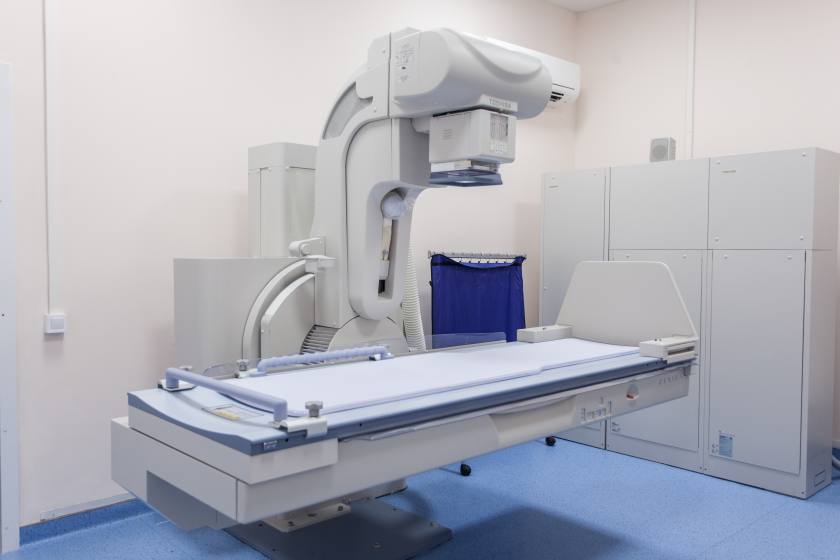
The device has a design that allows patients with claustrophobia not to feel discomfort. The equipment has a built-in noise reduction system, as well as a system in which the ultrashort echo allows you to examine areas of tissue previously inaccessible to MRI scans, such as the musculoskeletal system and lungs. A more sensitive magnet allows for high-quality and ultra-precise examination of the heart with minimal respiratory arrest.
The Toshiba Elan 1.5T MRI machine is the "gold" standard for MRI diagnostics for all leading medical centers around the world.
Cost of services
Indications for MRI diagnosis
MRI diagnosis makes it possible to detect pathologies of the brain and spinal cord, namely various types of tumors, developmental pathologies, aneurysms, multiple sclerosis, as well as to determine the state of the brain after injuries and strokes. Magnetic resonance imaging is the main technology of examination in such diseases of the joints and spine as herniated disc, arthritis and osteoarthritis, joint injuries. With the help of MRI diagnosis you can detect tumors in soft tissues and internal organs; MRI, along with mammography and ultrasound, is used to detect breast cancer.
Contrast-enhanced MRI is mandatory before planning surgery, as it allows you to determine the feasibility of surgery, and also allows you to give an idea of the size and exact location of tumors and obtain information about the close proximity to other tissues. In addition, contrast-enhanced MRI is used in traumatology to assess the extent of joint damage from blows and injuries.
Contraindications for MRI
Diagnosis with MR tomography is contraindicated for people with various surgical steel implants in the body, the presence of titanium implants is not a contraindication, pacemakers and any electromechanical devices such as insulin pumps, as well as the presence of metal objects in the head and eyes. Also contraindicated is the presence of large tattoos on the body using metallized pigments.
Contrast-enhanced MRI has a number of contraindications. Do not administer the contrast agent to patients:
- with severe kidney and liver disease in the stage of decompensation,
- with a history of kidney or liver transplantation,
- with detected allergic reactions to contrast during previous studies.
It is also not recommended to conduct research during the first trimester of pregnancy, during lactation the procedure is possible, but it is necessary to refrain from feeding for the next 24 hours.
The expediency of MRI with contrast in patients with bronchial asthma, myeloma and severe cardiovascular failure is decided by the doctor.

Preparation for MRI of the fetus and placenta:
MRI of the fetus and placenta in the medical network "Welfare" is performed by experienced and qualified specialists on MR tomographs of expert class (TOSHIBA Vantage Titan 1.5T) with a large tunnel diameter (71 cm) and a load capacity of the scanning table (up to 200 kg). Due to these characteristics, the device allows you to comfortably place the patient during the examination, even in late pregnancy and provides comfort even in the presence of claustrophobia.
Position of the patient at inspection: lying on a back. If it is difficult for a pregnant woman to lie on her back, the examination is performed in a supine position on the left side. The examination is performed with free breathing (without delay). The duration of the procedure is 30 minutes. If desired, the child's father may be present during the procedure.
Preparation:
- The procedure is most often performed in the morning, when the mother and fetus are at rest.
- The last meal is four hours before the examination.
- Caffeinated beverages should not be consumed on the day of the examination.
- Immediately before the procedure it is necessary to empty the bladder.
Contraindications for MRI:
- The presence in the body of electronic medical devices (pacemakers and others);
- The presence of metal elements in the studyareas (prostheses, clips, rolling pins);
- Restless behavior (panic attack, psychomotor agitation), which is diagnosed;
- Inability to maintain immobility during the examination (for example, due to severe pain);
- The need for permanent resuscitation (artificial respiration, etc.);
- The need for constant monitoring of vital signs (ECG, blood pressure, respiratory rate, etc.).
- Despite the fact that MRI is a safe method of diagnosis, it is not recommended in early pregnancy (up to 18 weeks). The optimal time is from 24 weeks, when the most clear and detailed visualization of all organs and systems of the fetus is possible, which will avoid diagnostic errors.
MRI of the fetus and placenta significantly improves the prognosis of successful childbirth and subsequent treatment of abnormalities.


%402x.png)
%402x.png)