Brain electroencephalography (EEG) is a noninvasive diagnostic technique for assessment of functioning and state of the brain. The assessment is performed through the registration of electrical pulses read by sensors on the scalp.
Electroencephalography is a highly sensitive diagnostic technique that takes into account the minimal effect of the stimulation pulses and following changes in the brain, and also registers frequency oscillations of electrical pulses in milliseconds. A doctor can ask a patient to wink, and also can use sources of noise and light to magnify the oscillations to receive these data.
EEG is a diagnostic procedure that allows to reveal neurological diseases and pathologies of brain activity. This examination method does not affect a patient and can be used also for children.
The following complains of a patient are indications for EEG:
- frequent headaches;
- insomnia, poor sleep;
- dizziness, syncopes;
- epileptic seizures.
All these symptoms can be the result of poor brain circulation, neoformations or even irreversible processes in the brain. Besides, they can result from abnormalities in the nervous system.
EEG is mandatory for the people that had traumatic brain injuries and neurosurgeries. It is a mandatory diagnostic technique for patients with epilepsy.
The procedure is absolutely safe and has no contraindications
The only obstacle for it may be the presence of injuries, sutures, bandages and signs of infectious processes on the surface of the head. In such a case, diagnostics is performed only after the wounds are healed.

Cost of EEG services
Description of the procedure

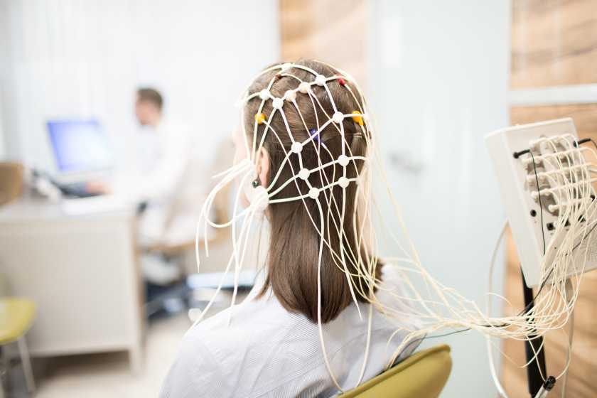
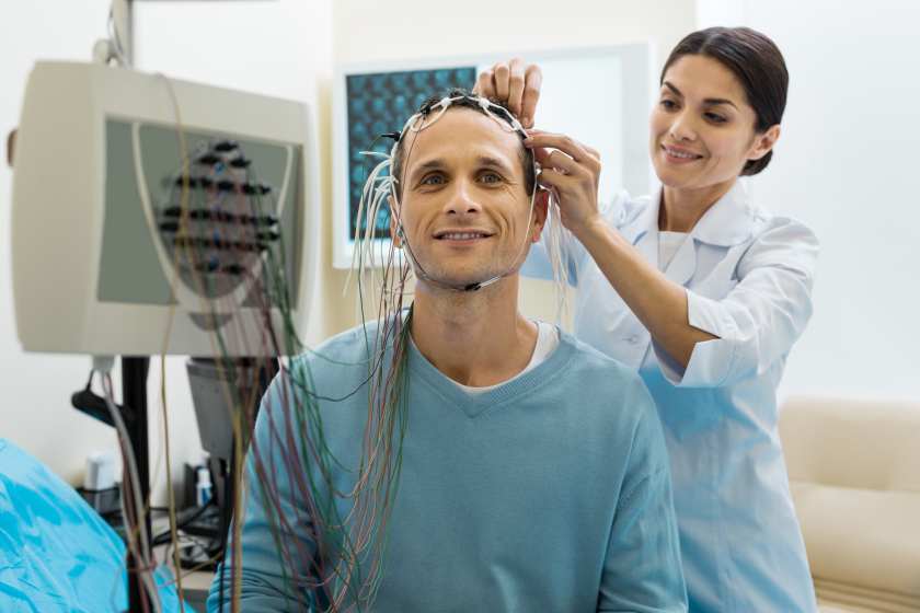
Brain activity is registered using a special device, encephalograph, that consists of three parts:
a cap with electrodes and wires, a unit that magnifies and reads the pulses and a display for presentation of the results.
An encephalograph is located in a small room closed off from noises and light where the examination takes place.
The examination may be performed in sitting position or lying on a couch. A patient wears a cap with wires on his or her head, turn on an encephalograph, and the device begins to read and transmit the information on the display in the form of a graph.
Electrical field power and its distribution by different brain regions can be fixed in this period. The so-called functional tests aimed at magnifying electrical pulses transmitted from the brain can be used during the procedure. The patient can be asked to wink quickly, to look at the light, to listen to harsh sounds, to change breathing rate and depth. Functional tests allow to reveal hidden functional changes in the brain.
The doctor takes off the electrodes after the procedure, prints the results and gives them to the patient.
The duration of this diagnostic technique depends on the region to be examined and the number of functional tests in the monitoring. The procedure takes of about 30 minutes at the average.
Preparation
To perform an electroencephalography, it is necessary to carry out a series of preparatory activities as follows:
- Discontinue anti-seizure, sedating and anxiety medications 72 hours before the procedure (if it is possible).
- Do not drink coffee and energy drinks, do not smoke 24 hours before the procedure.
- Wash your hear with shampoo the day before the procedure. Do not use foaming cremes, hair lacquers, mousses and other styling products.
- It is recommended not to be hungry before the procedure.
- Take off all the metallic accessories (earrings, necklace, clips, hairpins), let hair down before the examination.
It is important to stay calm and be ready for noises and flashes of light during the procedure and try not to react.
Read more about EEG in children and adults
Our advantages
Our team
Choose the nearest clinic
ISO certificates


Accreditation certificates


Medical practice licenses


%402x.png)
%402x.png)
