Echocardiography
Echocardiography or the heart ultrasound is the fastest and painless way to receive the information to prevent and treat cardiac diseases. During echocardiography a doctor can assess the state of the heart and the major vessels, to reveal pathological changes in the valvular heart apparatus, to diagnose pathological changes in the myocardium anatomy, to monitor the sanguimotion.
Echocardiography allows cardiologists to diagnose most of the cardiac diseases at their early stages quickly and precisely.
Echocardiography shows the following conditions:
- backflow on the valves and the state of the valves;
- dimensions of cardiac chambers and valves;
- heart tumors;
- cardiac malformations (congenital or acquired);
- heart functioning abnormality of professional athletes;
- cardiomyopathies;
- results of surgeries in cases of cardiac malformations.
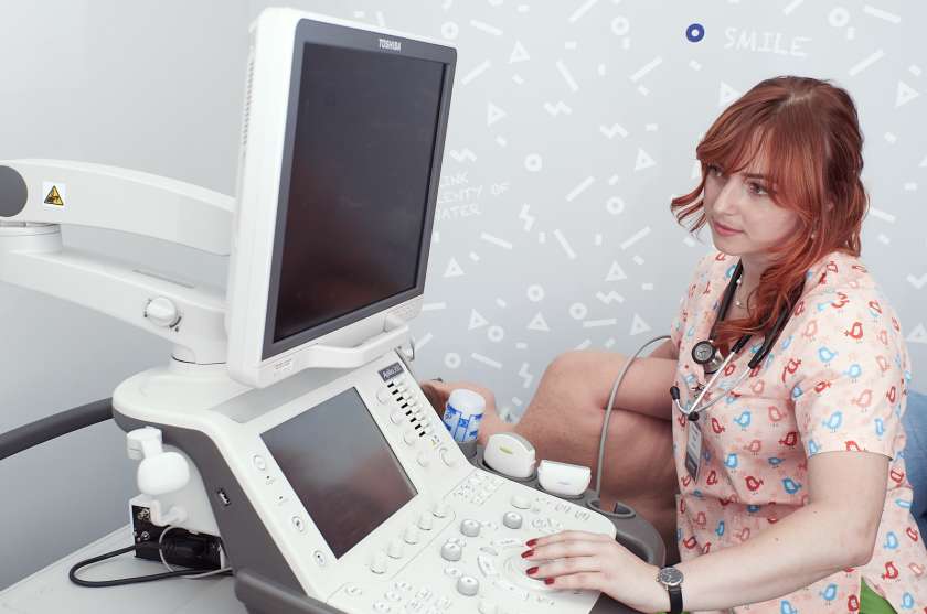
Description of the procedure:
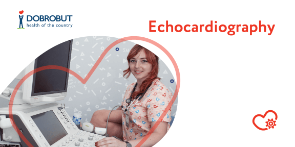
A patient has to strip to the waist, take off the accessories and lie on a couch. A doctor puts the heart rate sensor on certain points of the chest. The sensor transmits the ultrasound to the powerful computer that transforms waves into an image. A cardiologist sees a movable heart image during systole and diastole in real time.
The examination lasts 30 minutes, the patient can receive the report just after the examination.
Heart ultrasound is performed by an ultrasound specialist or a cardiologist. There are no absolute contraindications for this type of the examination.
There is one more echocardiography technique, the transesophageal echocardiography, when a special sensor is inserted that allows examining the patient's heart from different perspectives to reveal clots or to specify a diagnosis.
Such an echocardiography requires the following preparation:
- do not eat within 6 hours before the examination;
- take off your dental appliances before the examination if any.
A patient receives injections of the specialized medications that make the procedure more bearable.
This heart ultrasound technique has certain contraindications:
- esophageal pathology;
- osteochondrosis of cervical spine;
- diaphragmatic hernia.
Our advantages
Our servises
Choose the nearest clinic
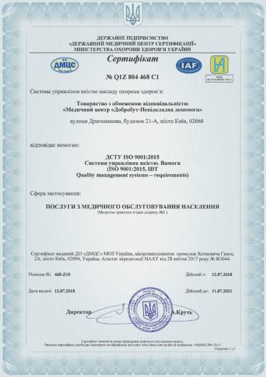
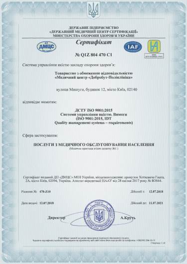
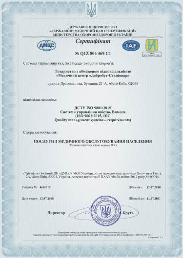
ISO certificates
Accreditation certificates
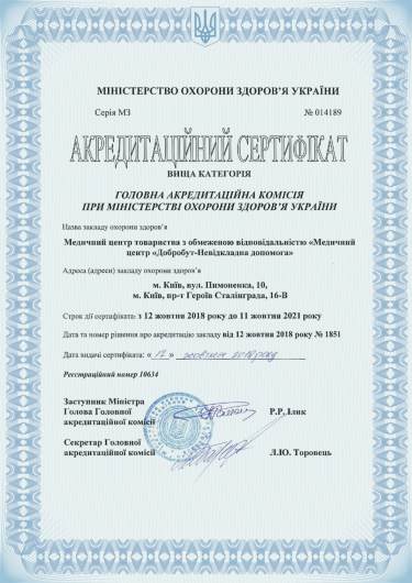
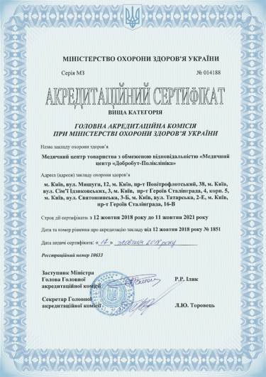
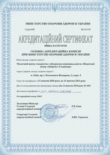
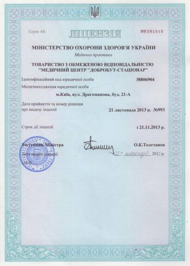
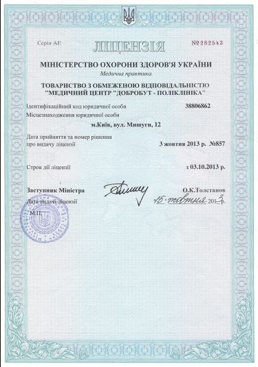
Medical practice licenses
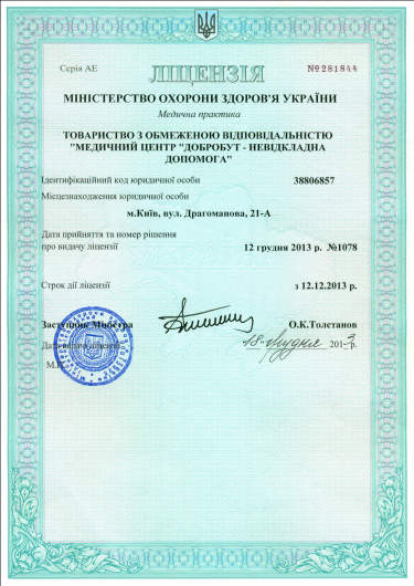








%402x.png)
%402x.png)
