CT of thoracic organs
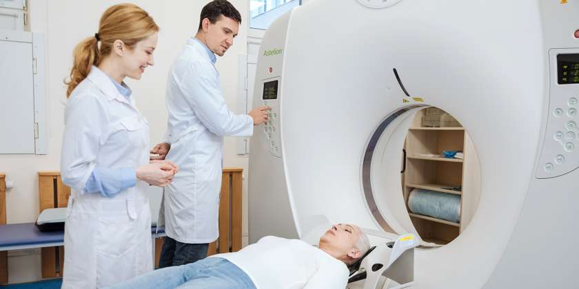
Computerized tomography of thoracic cavity is one of the most informative techniques for visualization of ribs, sternum and mediastinum organs. It is recognized as the “gold standard” of radiological study.
The principle of the method
This type of diagnostics is performed with the use of high-technology devices, tomographs. The method is based on the ability of X-rays to reduce their intensity when they pass through tissues. In the result of the computed processing high-quality, clear layer-by-layer images of the region of interest are received that allow for diagnosing of pathologies of bronchial tubes, lungs and heart, change in lymph nodes, vessels, esophagus and osseous structures.
Computerized tomography of lungs and other thoracic organs may be traditional (step-by-step) and spiral (single-slice and multi-slice). In the first case, scanning of one tomographic slice is performed, in the second case, the data from the whole organ being examined are received. Spiral CT allows to reconstruct and to take a close look at up to 640 slices.
When the computerized tomography of lungs is prescribed, who has contraindications for such type of diagnostics
There are the following indications for the lung CT:
- chest injuries;
- preparing for a surgical intervention;
- respiratory or cardiovascular system malformations;
- suspected presence of liquid or commissures in the pleural cavity;
- benign and malignant neoformations;
- diseases of infectious inflammatory genesis;
- lymphadenopathy of an undetermined origin;
- explicitation on the presence and localization of destruction cavity focalities or seeding;
- clarification of a diagnosis in case of detected shadows during an X-ray radiography procedure;
- necessity of assessment of the respiratory organs condition in case of the pleural fluid presence.
In case of an oncologic process, the computerized tomography of lungs in Kyiv will help:
- to determine the precise localization and dimensions of tumors in thoracic or mediastinum organs;
- to determine hidden metastases;
- to exclude a recurrence of the tumor growth after a surgical treatment;
- to assess the degree of spread of the tumor to adjacent areas.
In some cases, diagnostics may be performed even on the patients put on mechanical ventilation.
Lung CT in Kyiv has the following contraindications:
- pregnancy;
- allergy to contrast agents;
- some mental disorders;
- muscle spasms and cramps;
- pancreatic diabetes and renal disease.
What a patient needs to know about the process of CT of thoracic cavity
Before the beginning of the examination, a specialist talks to a patient in order to:
- make certain of the absence of contraindications to CT;
- tell a patient about the course of the procedure;
- get the hard copies of previous examinations (if there are any).
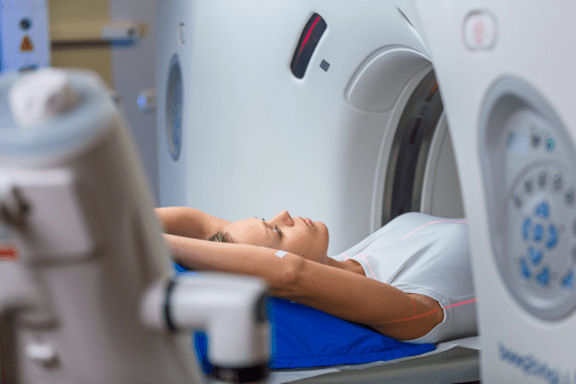
After that, the patient’s body is fixed on a special table and moved to the tomograph camera. The procedure lasts of about half an hour.
The price for computerized tomography of lungs is relatively low, so this type of diagnostics is available to everybody who has indications to it.
Important note. If you feel worse or have a panic attack during the procedure, please let the medical staff know.
Remember that you should not eat and drink for hours before a CT of thoracic organs with contrast agent.
CT of thoracic organs is an important diagnostic procedure, so we advice to undergo it only in a professional facility.
Article author: Andriy Ratushnyuk
Publication date: 19.12.2020
Bibliography
- Rouprêt M, Babjuk M, Compérat E, et al. EAU Guidelines on Urothelial Carcinomas of the Upper Urinary Tract. In: EAU Guidelines. Edition presented at the EAU Annual Congress Stockholm 2014. ISBN 978-90- 79754-65-6.
- Русаков И.Г., Быстрое А.А. Хирургическое лечение, химио- и иммунотерапия больных поверхностным раком мочевого пузыря. Практическая онкология № 4, 2003; С. 214-24.
- Мартов А.Г., Ергаков Д.В., Байков Н.А., Поминальная В.М., Соломатов И.А. Трансуретральное удаление опухолей мочевого пузыря единым блоком // Онкоурология. 2015. Т. 1, № 1. С. 41–49.
More about Computerized tomography at Dobrobut
Our advantages
Our services
Our doctors
Choose the nearest clinic
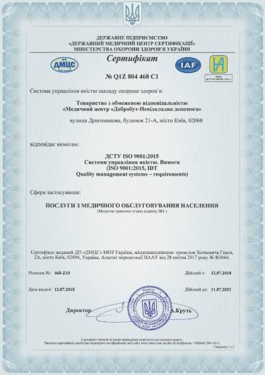
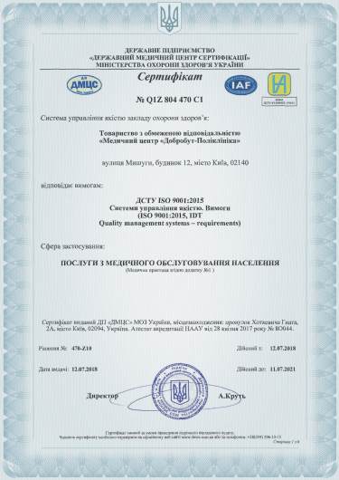
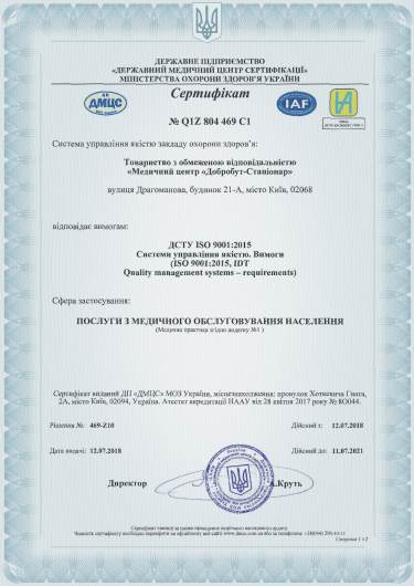
ISO certificates
Accreditation certificates
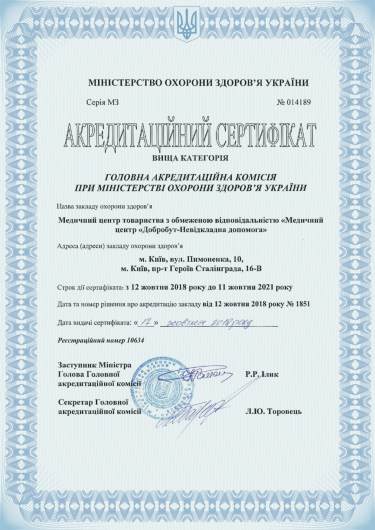
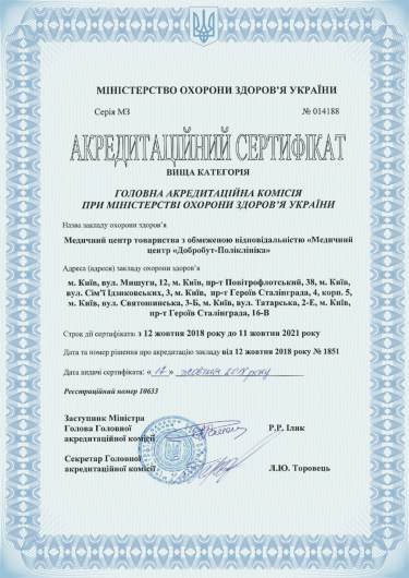
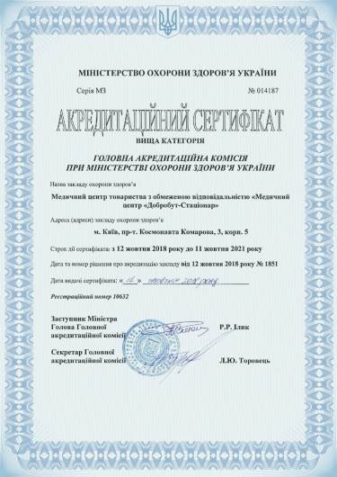
Medical practice licenses
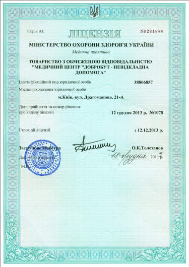





%402x.png)
%402x.png)
