CT scan
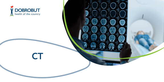
Computerized tomography is one of the modern diagnostic techniques characterized by fast examination speed, high accuracy and information value. It helps to receive the comprehensive information on the state of internal organs, bones and vessels. The method is of particular importance in the work of oncologists, as it allows to obtain a quality 3D image where the slightest changes in the structure of organs and tissues can be seen, to define the localization and density of the neoformation and to make an assumption about the tumor nature: if it is malignant or benign.
By contrast with a usual X-ray apparatus, the CT apparatus is able to make many X-ray scans simultaneously due to its construction, in particular, a tube and a set of detectors rotating about the patient at high speed. Thus, the method allows to get a layer-by-layer image of a particular human body area. Computerized tomography performed correctly precludes the probability that some processes in tissues and organs remain unnoticed.
Diagnostics with the usage of the CT scanner is performed in two ways:
- without a contrast agent;
- with the injection of a contrast agent before the procedure.
As CT is a radiological diagnostic approach, the investigative procedure is performed only at the direction of a clinical oncologist or a consulting physician. The consulting physician makes the decision about the way to perform the procedure.
A consultation by a radiotherapist to read the test results is of great importance too.
Equipment
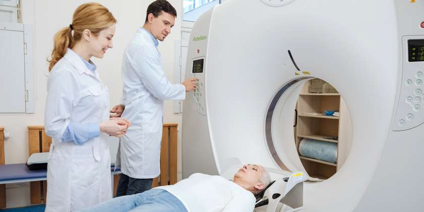
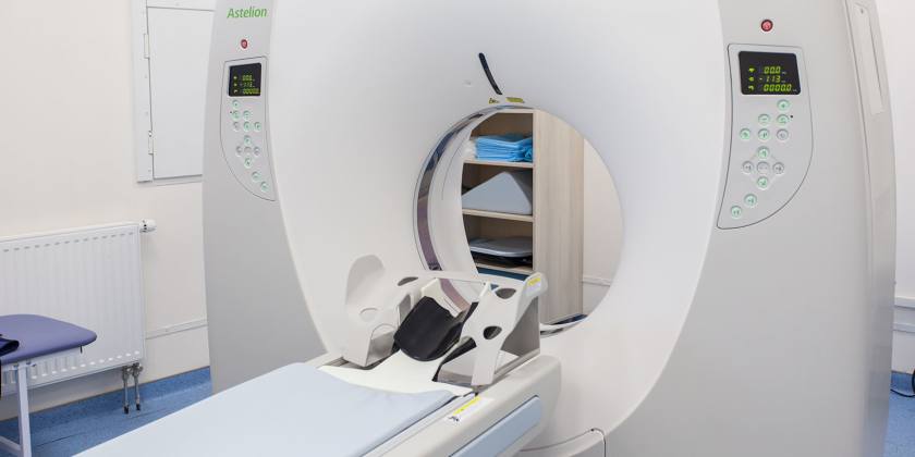
Dobrobut clinic chain uses the equipment by the leading medical devises manufacturer Toshiba, in particular, Astelion 16 row model, to perform the computerized tomography.
The CT scanner allows to carry out the 32-slice dynamic visualization of the organ at the minimum radiation exposure that is 4 times less than at usual X-ray examination. The image of the organ is converted into the digital format and allows to create its reconstruction in 3D format. The scanner allows to make 0.5 mm slices at a speed of 15 images per second. The equipment is used also for brain and heart examinations.
Astelion 16 row CT scanner is a modern equipment with advanced options that allows to perform the high-accuracy diagnostics and receive contrast images with the minimal period of time.
Description of CT procedure
Indications
Computerized tomography is more often used to clarify a diagnosis after X-ray or ultrasound. Examining various areas, including CT of the whole body, it is possible to reveal both various benign and oncological diseases (cancer detection), and also infectious, cardiovascular, respiratory diseases, surgical pathologies, etc.
The main indication for the computerized tomography is the necessity of detailing of the pathologies found with screening methods. The diagnostic method is effective in case of brain and spinal diseases, diseases of ENT organs, spinal chord, vessels, heart, lungs, liver, kidneys, pancreatic gland.
Injection of a contrast agent before the CT procedure is the most requested when examining blood vessels, lungs, abdominal organs.
Contraindications
Computerized tomography is not performed for women at any period of pregnancy, and also for the patients suffering from severe form of any disease.
Computerized tomography is prescribed for small children only if it is impossible to apply other examination methods or their results are insufficient.
The contrast agent used for the contrast-aided CT is a preparation based on iodine. Due to its possibility to accumulate in the body, it allows to receive more detailed and clear image during an examination.
The contraindications to the contrast-aided CT are as follows:
- Allergic reactions to iodine and iodine-containing preparations.
- Renal disease (iodine is metabolized and eliminated from the organism mainly by kidneys).
- Severe hepatic impairment.
- General grave condition of a sick person (for example, in case of severe pancreatic diabetes, asthma, erythroid myeloma, thyroid diseases).
- Decompensated thyroid diseases.
- Use of metformin or alternative drugs in case of pancreatic diabetes: to discontinue for 2 days before or 2 days after the examination.
Preparation
You should prepare yourself for computerized tomography.
- A light dinner in the evening before the procedure.
- A cleansing enema before going to bed of taking medications for evacuation of bowels (in case there are no contraindications).
Examination of abdominal organs is performed under fasting conditions.
You should drink 1–1.5 liters of natural water 1.5–2 hours before the procedure, 200 ml of it should be drunk just before the procedure.
Rectal examination is performed under pneumocolon conditions: the air is introduced into the rectum, that is why the thorough evacuation of bowels with Fortrans or similar medications is needed.
To perform the contrast-aided CT it is necessary to do blood chemistry for creatinine, and you should also mention allergic reactions to iodine and iodine-containing preparations in the past. If there are severe allergic reactions like Quincke's disease, anaphylaxis, the contrast-aided CT is forbidden and should be replaced with alternative diagnostic methods like MRI and ultrasound.
Description of CT procedure
During the procedure a patient is located on the special table to exclude the possibility of artefact appearance due to movement; it is important to stay still.
An intravenous catheter will be placed to inject the contrast agent.
The patient is monitored through the observation window during the procedure, and there is voice communication. Modern equipment allows a doctor to see structure, shape and dimensions of any pathological areas perfectly, zoom or rotate an image to obtain the most informative angles and dynamically assess the state of organs and tissues.
A procedure without contrasting lasts less than 15 minutes, with contrasting – up to 30–40 minutes.
Reading of the results
Ready images and the examination saved on a СD can be received 40 minutes after the procedure. Besides the results, no less important is to obtain their detailed reading and interpretation of the image by highly qualified radiologists with reach work experience with such an equipment and extensive medical knowledge. Usually, the description takes not less than 2 hours, in particular complicated cases the results can be issued the next day.
Our advantages
Our services
Our doctors
Choose the nearest clinic
ISO certificates
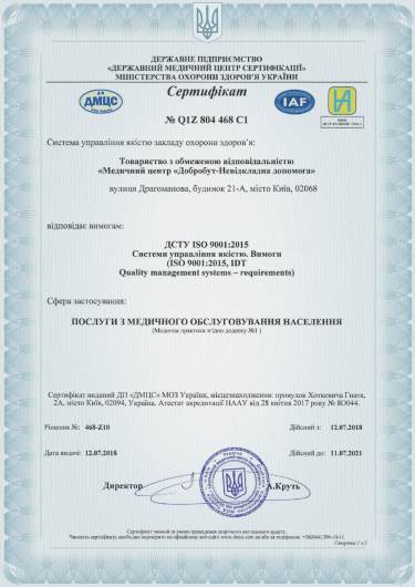
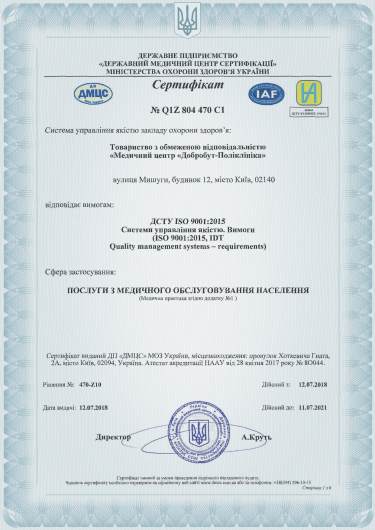
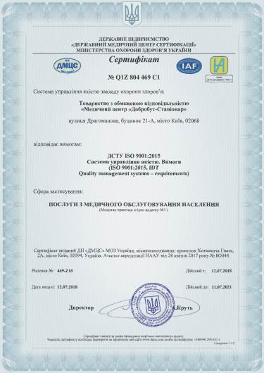
Accreditation certificates
Medical practice licenses









%402x.png)
%402x.png)
