Rectoromanoscopy
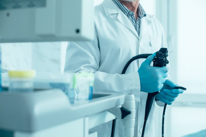
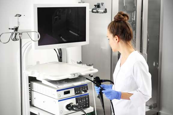
Rectoromanoscopy is a method of instrumental visual examination of the rectum and distal sigmoid colon by a rectoscope introduced into the anus. Rectoromanoscopy is the examination method prescribed to a patient consulting a proctologist with complaints of bleeding, mucus and pus discharge from the anus.
Procedure
Rectoromanoscopy is performed in the knee-elbow position of the patient. The doctor lubricates the end of the tube with petroleum jelly and inserts the rectoromanscope tube into the rectum carefully moving it to the depth of 20-35 cm. To straighten the walls of the rectum for better viewing, by means of a double pump, a little bit of air is pumped into the rectum. This method along with other endoscopic examination techniques allows collecting material for histologic research (biopsy).
Indications for rectoromanoscopy
- pain in the anus,
- discharge of blood, mucus or pus from the anus,
- stool disorders (constipation, diarrhea),
- suspected rectum and sigmoid colon diseases
Rectoromanoscopy for prophylactic purposes is becoming very popular. As a preventive measure for early diagnosis of malignant neoplasms of the rectum it is recommended once a year for people over 30 years. Rectoromanoscopy should be preceded by X-ray examination of the colon, because minor changes in the rectum (small tumors, infiltration processes or proctitis) are diagnosed only by endoscopic examination.
Contraindication
- cardiac decompensation,
- severe general condition,
- narrowing of the lumen of the anal valve and rectum,
- acute inflammatory processes in the anus (acute paraproctitis, thrombosis of hemorrhoids),
- stenotic tumors of the anal canal;
- chemical and thermal burns in the acute stage.
Preparation for rectoromanoscopy
Thorough cleansing of the colon from the contents is an important condition for rectoromanoscopy. The day before rectoscopy patients are prescribed a slag-free diet during the day with only tea in the evening. Examination is conducted on an empty stomach. In the evening and two hours before the examination the intestines are cleansed with enema. Before rectoromanoscopy, make sure to assess the anus condition by finger rectal examination. This allows easily detecting anal eczema, dermatitis, warts, external fistula openings, perianal thrombosis, skin ulcers (marigolds), protruding hemorrhoids or polyps, pararectal abscesses, rectal prolapse and tumors.
Two options to get prepared for examination:
- Option 1: the day before - slag-free diet (diet No 4 - exclude meat, vegetables, fruits), in the evening - a light dinner, after 6.00 pm no meal, only tea. Make 2 cleansing enemas the night before, at 9.00 pm and 10.00 pm; on the day of the examination: 1 enema in the morning and 1 enema two hours before the examination. It is allowed to use NormacoI instead of enemas.
- Option 2: the night before - a light dinner, after 6.00 pm no meal, only tea, take 2-3 packets of Fortrans (1 packet per 20 kg of body weight) from 5.00 pm to 9.00 pm (1 sachet dissolved in 1 liter of still water, drink within an hour).
The examination lasts 30 minutes. The doctor’s opinion is provided within 15 minutes after the end of the examination.
Cost of services
Our advantages
Service packages
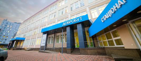
Our clinics
ISO certificates
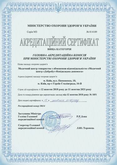
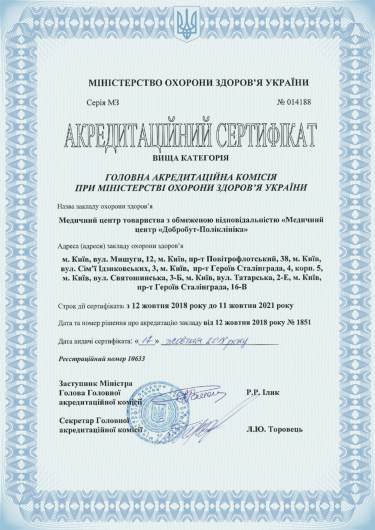
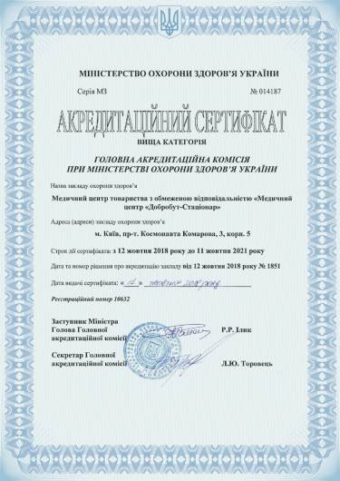
Accreditation certificates
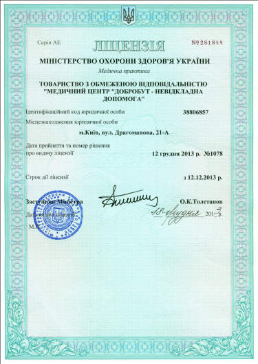
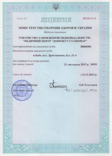
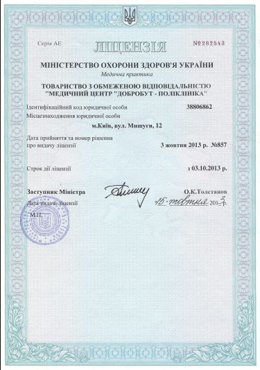
Medical practice licenses


%402x.png)
%402x.png)
