Our directions
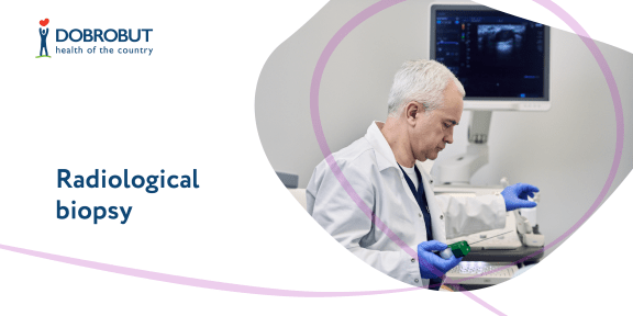
Radiological biopsy
Radiological biopsy
Additional visualization techniques such as CT and ultrasound are used for better targeting when taking the material from the most suspected area for examination during the biopsy. The choice of the technique depends on which of them provides better visualization of the area.
After doctors have received a tissue sample, it is sent to a pathologist for examination; the latter analyses the sample received under the microscope and determines if the removed tissue is malignant or benign.
Examination results contain a detailed description of the tumor type. Additional immunohistochemical and/or molecular tests can be performed to determine the type of the tumor or its characteristics more precisely and to choose the treatment approach.
Bibliography
- Abul-Kasim K. et al. Intradural spinal tumors: current classification and MRI features. Neuroradiology 2008; 50: 4: 301—314.
- Cicero G, Giugno A, Maugeri R, Graziano F, Basile L, Iacopino DG. A rare case of extra-intramedullary dorsal tanicitic ependymoma, radically removed with intraoperative neurophysiological monitoring. Acta Medica Mediterranea. 2015;31:1229.
- Черкашин М.А. , Пучков Д.Д., Реут Е.А - Трепан-биопсии солидных опухолей под контролем компьютерной и магнитно-резонансной томографии - «РМЖ» №6 от 27.11.2019 стр. 3-9
- Burke A.P., Veinot J., Loire R. et al. (2004) Tumors of the heart: introduction. In: Travis W. (Ed.) Tumours of the lung, thymus, pleura and heart. Springer-Verlag, Lyon, p. 251–253.
Our advantages
High qualification
Wide range of medical care
Affordable prices
Flexible system of discounts for program services
24-hour-a-day work
The contact center informs and records patients 24/7
Radiological biopsy in Dobrobut
View all services
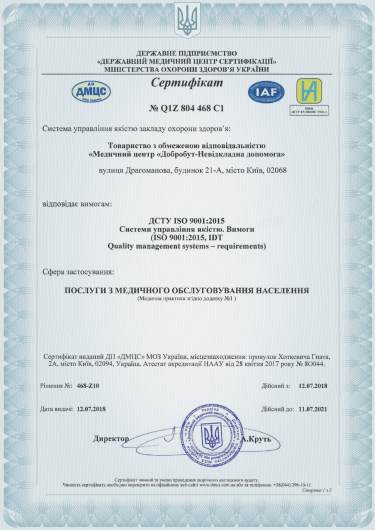
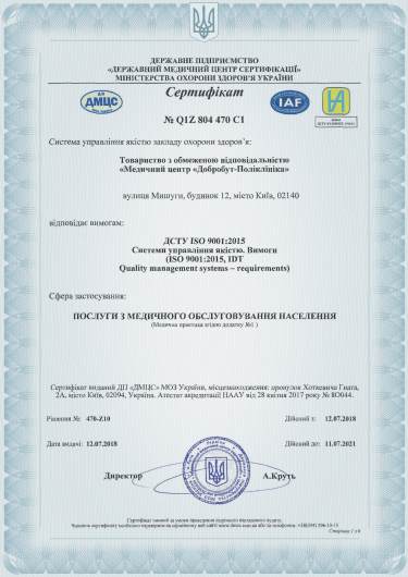
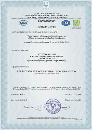
ISO certificates
of Dobrobut clinic chain
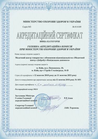
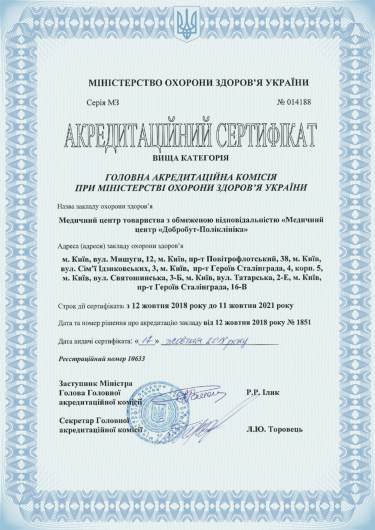
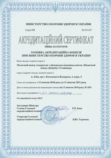
Accreditation certificates
of Dobrobut clinic chain
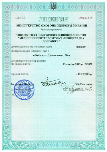
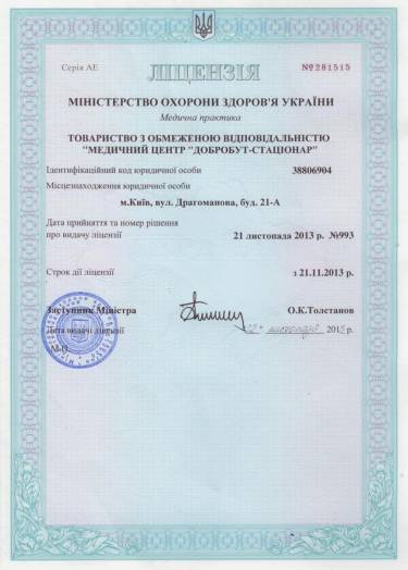
Medical practice licenses
of Dobrobut clinic chain
Terms of online services
Public contract for the provision of medical services
Terms of vaccination services
Patient verification
2012 - 2026 Dobrobut MN. All rights reserved


%402x.png)
%402x.png)
