Diagnosis in dermatology
Skin diseases can have various forms (chronic or acute), various manifestations and causes. That is why diagnostics of skin pathologies requires systemic and complex approach.
The advantage of skin diseases diagnostics is that the organ is on the surface and available for a visual check-up by a doctor. A doctor can often make assumptions on the character and causes of a disease, and determine a provisional diagnosis basing on shape, appearance, manifestations and location of pathologic areas.
Dermatologists use laboratory diagnostic techniques such as clinical blood analysis and blood chemistry, biopsy, allergic tests and other examinations to specify or confirm a diagnosis. One of the main instrumental diagnostic techniques is dermatoscopy.
Laboratory diagnostics
Laboratory diagnostics provides a dermatologist with the clinical data on the patient’s general health condition.
The following laboratory diagnostic techniques are used in dermatology:
- clinical blood analysis and blood chemistry;
- allergic tests;
- pathohistological examination of tissue samples after a biopsy;
- test for mycotic infections;
- bacterial examination.
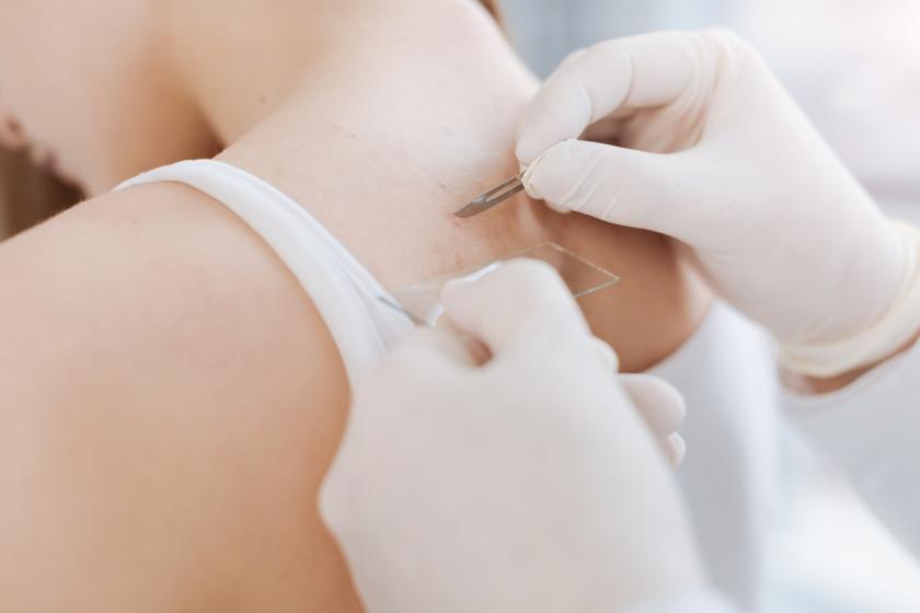
The biopsy is performed to take skin samples for a pathohistological examination: a tissue sample is taken from an abnormal skin area and sent for examination to a pathohistological laboratory. The examination plays an important role in diagnostics and early detection of skin oncological diseases.
A doctor makes a scraping to perform tests for a mycotic or another type of infection, and such diseases as itch or demodecosis, and sends the samples to a laboratory for examination.

Instrumental diagnostics
Dermatologists use various additional instrumental diagnostic techniques for more detailed check-up and diagnostics of skin neoformations.
The lamp emits ultraviolet (invisible part of the spectrum) that helps in diagnosing mycotic and bacterial infections, and enhances the visibility of pigmented lesions caused by various diseases, for example, vitiligo. The lamp is used in a dark room, its rays are targeted to the affected skin area.
Diascopy is the studying of skin coloration changes when pressing. During diagnostics, a doctor uses a special glass applied to the affected skin area and presses it. Various skin coloration changes may indicate various diseases. For example, in case of sarcoidosis the skin becomes olive brown.
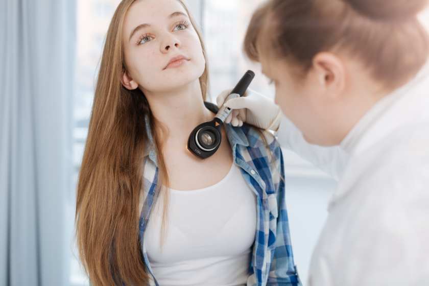
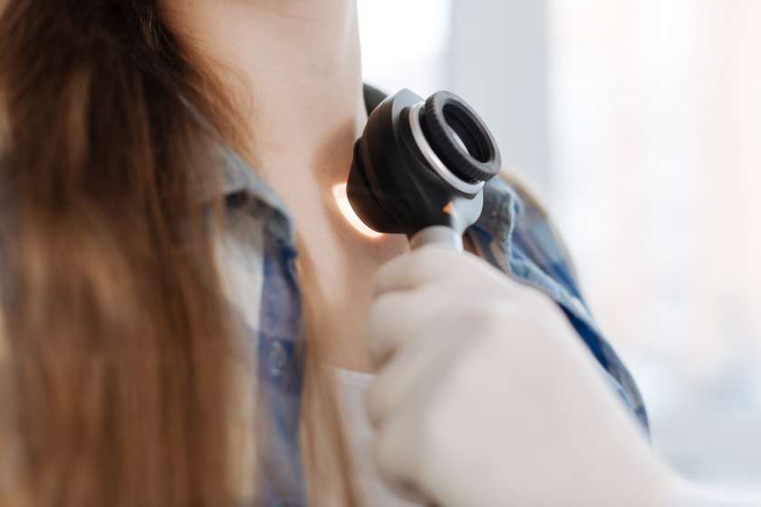
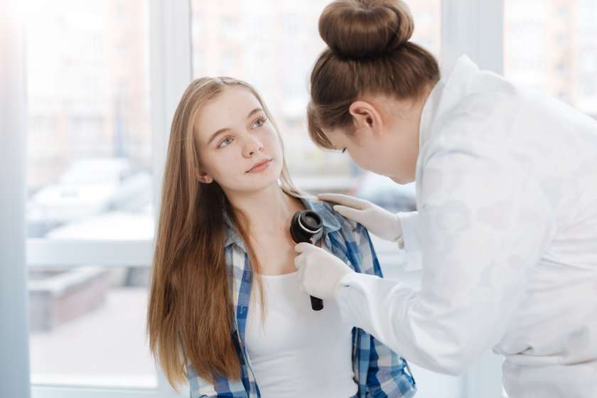
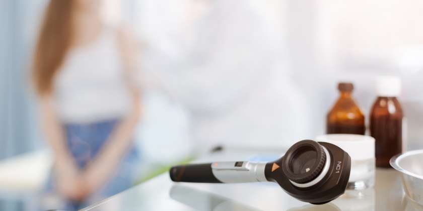
Dermatoscopy is the technique that allows determining of skin neoformation character. Diagnostics is performed with a special device, dermatoscope that magnifies the image by 10 times. With this device, a doctor can study the structure of the neoformation, its symmetry, edges, and extent of growing into skin, and to determine if it is benign or malignant. This absolutely safe and simple diagnostic technique allows detecting and removing of the melanoma (skin cancer) at the earliest stages.
Dermatoscopy is necessary in the following cases:
- many new moles came up or begin to come up on the skin;
- the mole increased in size, changed its shape, color or there are any other changes in its appearance;
- itching in the mole area. Sloughing or any other type of discomfort and feeling of uneasiness;
- removal of a skin neoformation is to occur;
- you have had sunburns or you often use a tanning room;
- many pigment stains and raised formations (birthmarks) with the diameter of more than 5 mm.
Risk factors for the developing of skin cancer are:
- Fair hair and skin, gray, green or blue eyes;
- Excessive exposure to the sun or staying in a tanning room;
- Close relatives with oncological diseases, skin cancer included.
If you have no complaints, you should undergo dermatoscopy 1–2 times a year, and 3–4 times a year if you are over 60 years old.
Preparation
The procedure does not require any preparation, cosmetic products (cream, body milk, shower gel, etc.) on the skin are not the contraindications for dermatoscopy and do not affect the reliability of the results.
Detailed on diagnostics at Dobrobut:
Our services
Our clinics


ISO certificates
Accreditation certificates
Medical practice licenses
%402x.png)
%402x.png)