X-ray diagnosis
X-ray diagnosis is examination of any part of the body with X-rays to obtain a fixed image on a sensitive film or screen.
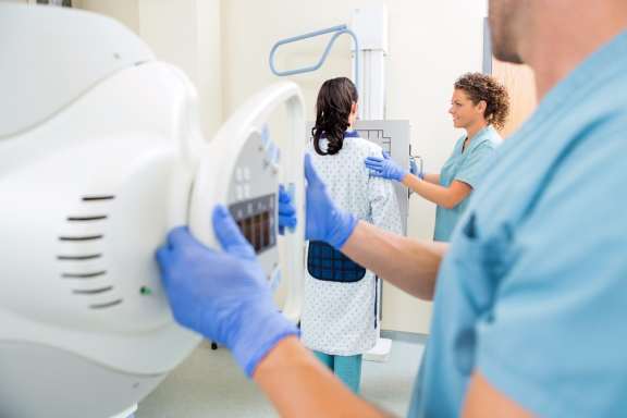
The X-ray technician enters the patient's data into the computer. The patient stands motionless in front of the screen for a moment. The doctor in the next office (control room) sees the image of the lungs on the display and announces the diagnosis. Formerly you had to wait for the image several hours, now it is ready only in 10-15 seconds. This is how a digital X-ray unit works today.
What other advantages does medical equipment have? Is it possible to make X-ray for small children? How often can you be examined to detect pathology at an early stage?
Modern digital X-ray equipment RADSpeed developed by Japanese scientists and engineers and manufactured by MF SHIMADZU CORPORATION in 2009 allows making X-ray without harm to health up to 6 times a year, because during the examination a patient receives the radiation dose of 10-15 times less than with older X-ray units.
The advantage is that you do not need to buy the film and spend time on its development. Previously, 10-12 images out of 100 were of poor quality: either the laboratory assistant kept it too long in the solution, or the developer was old, or the solution was cold or the film was defective. And the patient was referred for the re-examination, thus for another irradiation dose. Now this is practically an exception. The computer allows directing light to a particular area, enlarging it by 5-10 times or making a positive: the human eye better recognizes pathology in the positive than in the negative. In difficult cases, the image can be sent via the Internet to a medical specialist in any other health facility for additional consultation.
What diseases are detected by X-ray?
These are pneumonia, bronchitis, pleurisy, tuberculosis, lung cancer. The images show very well the skeletal system of the chest, heart disease and cardiovascular disease. At least ten people out of 100 are referred to a cardiologist or to a cardiac surgeon after X-ray examination. The X-ray image will help a gastroenterologist to diagnose the diaphragmatic hernia, the pediatrician - the curvature of the spine. The endocrinologist will get an answer as to whether this is thyroid or thymic disease. A digital device allows a radiologist to see thickening of 2-3 mm and diagnose tumor at an early stage. The X-ray unit allows using a contrast medium for examining organs of the urinary system (urography, urinary cystography), digestive organs (irrigography for the large and small intestine).
Use of the older X-ray unit was allowed for children after 14 years. The digital device reduces the radiation load. Besides, only one millisecond is enough to receive a quality image for a thin child. All you need to do is to persuade the child to remain still, not to cry and stay on the X-ray unit platform.
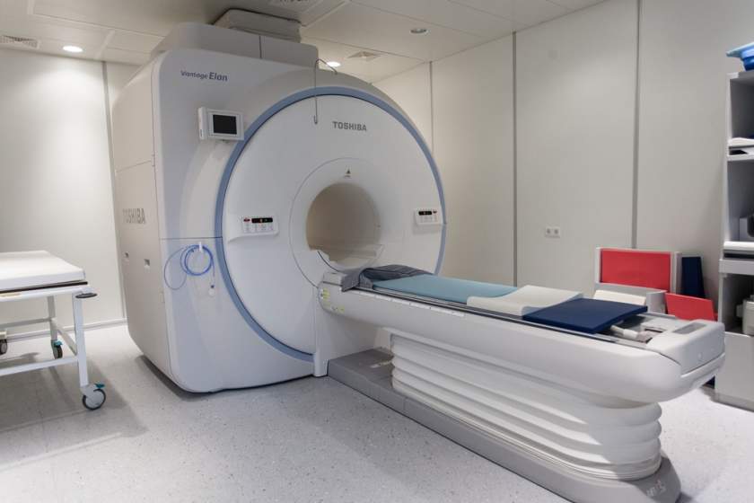
The modern equipment of an X-ray room allows:
- assessing structure of soft and dense tissue, for example, it is possible to detect scoliosis when inspecting respiratory organs) at the initial examination;
- significantly enlarging the image and see the structural changes of organs and tissues;
- making diagnosis as quick as possible due to shorter exposure (0.01-0.4 sec), which is safe for a patient;
- enlarging the size of specific areas, changing the contrast and brightness of the image.
Digital image processing of the modern X-ray unit can significantly improve the quality of images and improve the diagnostic value of the examination. This is very important when examining young patients who cannot hold their breath or remain still.
Saving the image on digital media allows comparing images over time (in case of chronic processes), making the required number of copies, selecting, enlarging and printing individual areas of the image.
All types of X-ray examinations are performed, including contrast and functional examinations.
X-ray examinations are performed only by a written referral from a doctor of the Dobrobut Medical Network or other health facility. It should be certified by the seal of a doctor or the health facility. To book for the X-ray examination, please, call the Dobrobut MN Contact Center.
More about radiography and radioscopy at Dobrobut
Cost of services
Our clinics
ISO certificates


Accreditation certificates

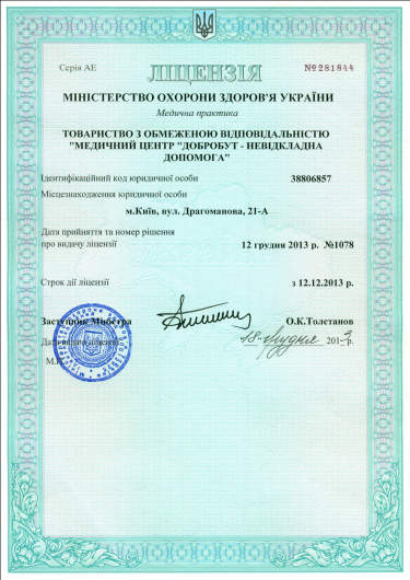
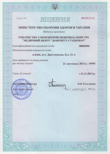
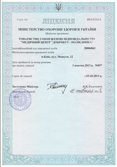
Medical practice licenses


%402x.png)
%402x.png)
