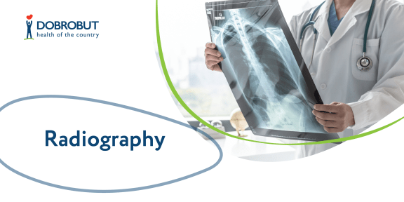
X-ray radiography is the first noninvasive diagnostic technique available to the humanity. Since its inception it has been updated significantly, more modern and safer X-ray machines appeared, but its operation concept has not changed. Organs and tissues are examined with X-rays, and the projected 2D image is plotted on the special film or paper.
X-ray radiography is the fastest and the simplest method to perform that allows to see changes in tissue structure of the examined organ and in thus to make a conclusion about its condition and the presence of eventual diseases.
Lung X-ray is the most common screening method of diagnostics that is recommended to everybody once in three years. Every so often X-ray radiography gives the information on a suspected disease that becomes a signal and a reason for further more detailed diagnostics.
Our advantages
Description of the procedure
X-ray radiography is prescribed in order to reveal the specific impairments of thoracic (lungs, heart), abdominal (liver, stomach, esophagus, duodenum and rectum), small pelvis organs (kidneys), and also joints and spinal chord.
X-ray is efficient for revealing of pneumonia, arthritis and myocarditis in case of infectious etiology of the disease.
X-ray radiography has no absolute contraindications. However, taking into account a low X-ray exposure during the procedure, the X-ray radiography is not recommended to undergo too often, and it is also prescribed for pregnant women, breastfeeding mothers and children under 14 years of age with care and only when it is absolutely necessary.
Modern X-ray machines allow to perform the procedure both in upright and in lying position. You should strip to the waist for the chest examination. You should take out all the metallic objects and take off the accessories before the X-ray radiography.
A diagnostician will help to find a required body position to receive the necessary projection of the organ being examined, after that you should stay still throughout the whole procedure.
The organs not participating in the diagnostics are covered with a protective lead apron, a doctor goes to another room and monitors the procedure from there.
Images are obtained in several minutes, all the procedure from the moment of entering to the room and preparation to the procedure will take less than 15 minutes. You can obtain the examination results in 10–15 minutes.
Specific preparation is not required for X-ray radiography. It is recommended to cut out gas-forming food several days before the scheduled examination of stomach and bowels, and to do a cleansing enema just before the procedure. X-ray examination of these organs is performed under fasting conditions.
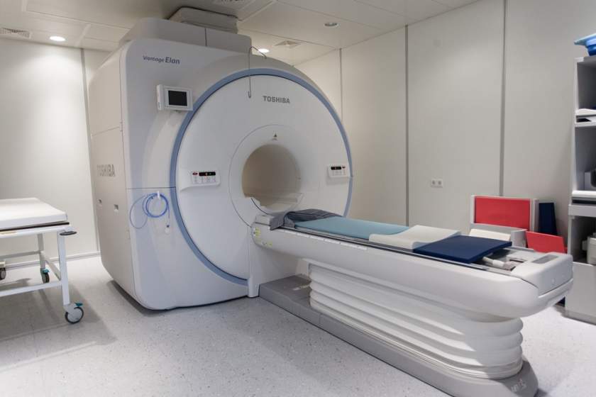
Cost of services
Choose the nearest clinic
ISO certificates
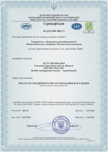
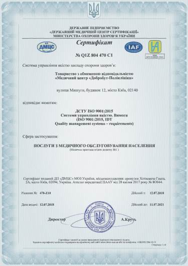
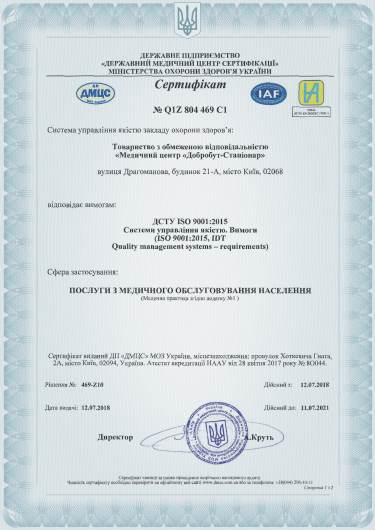
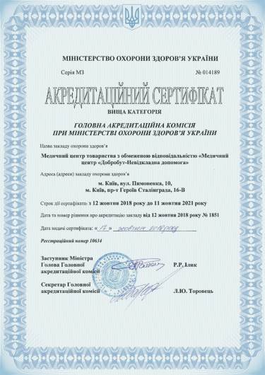
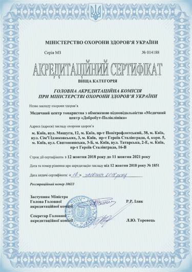
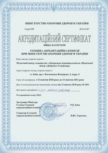
Accreditation certificates
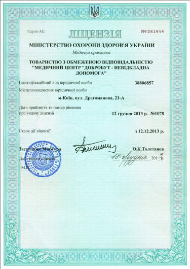
Medical practice licenses





%402x.png)
%402x.png)
